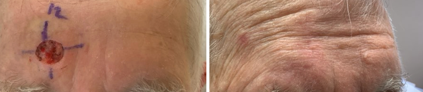
This patient had a surgical defect located directly over the right eyebrow. Reconstruction was performed using an O-to-L advancement flap, a technique chosen specifically to optimize both functional and cosmetic outcomes in this anatomically sensitive area. The skin adjacent to the defect was carefully mobilized and advanced into the wound, allowing closure with tissue that closely matched the surrounding skin in color, thickness, and texture.
This repair method was selected to minimize tension on the eyebrow and surrounding structures. Direct linear closure in this location could have resulted in upward displacement or distortion of the eyebrow due to vertical tension vectors. By utilizing an O-to-L advancement flap, tension was redistributed more favorably, helping to preserve the natural position, contour, and symmetry of the eyebrow while achieving a secure wound closure. This approach also helps minimize the risk of functional impairment and provides a more aesthetically pleasing final result.
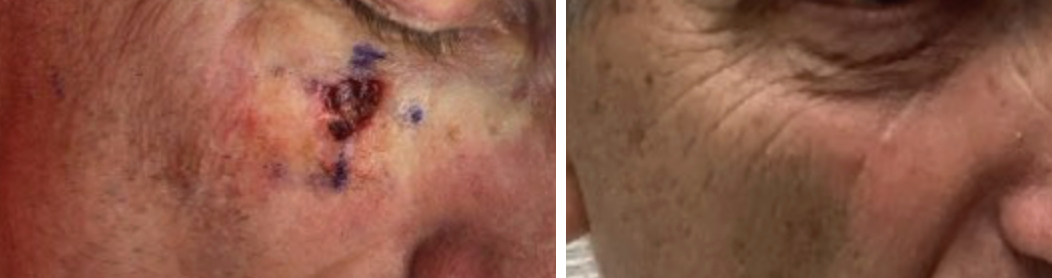
Defects in close proximity to the lower eyelid can be challenging to reconstruct because excessive tension may lead to distortion of the eyelid margin, ectropion, or other functional and cosmetic complications. In this case, the defect was amenable to a linear repair, allowing for a straightforward reconstruction while still preserving normal eyelid anatomy.
The closure was carefully designed and oriented to direct tension away from the lower eyelid. By angling the repair along favorable skin tension lines and avoiding vertical traction on the lid margin, the reconstruction minimized the risk of downward pulling or distortion of the eyelid. This approach provided excellent wound closure while maintaining the natural contour, position, and function of the lower eyelid, resulting in a favorable functional and aesthetic outcome.

This defect was repaired with a linear repair that was carefully designed and oriented to minimize tension on the lower eyelid. By strategically angling the repair, tension was redirected away from the eyelid, helping to preserve its natural position and function.
In addition to protecting the lower eyelid, the orientation of the closure allowed the final scar to be placed along favorable skin tension lines, improving cosmetic camouflage and reducing scar visibility. This approach provided a simple yet effective reconstruction, achieving secure wound closure while optimizing both functional preservation and aesthetic outcome.

Even minor distortion vermilion border of the lip may be readily noticeable and can affect both cosmetic appearance and lip function. Precise restoration of the vermilion border is essential to maintain the natural contour, symmetry, and appearance of the lip.
In this case, reconstruction was performed using a V-to-Y advancement flap. This technique allows adjacent lip tissue to be advanced into the defect while preserving tissue color, texture, and thickness match. The flap was specifically selected to maintain proper alignment of the vermilion border and minimize distortion of the surrounding lip architecture. By recruiting local tissue and carefully redistributing tension, the repair preserved the natural shape and function of the lip while achieving an excellent cosmetic outcome. The V-to-Y advancement flap is particularly well suited for defects in this area because it provides reliable tissue movement without creating excessive tension or significant alteration of the lip contour.
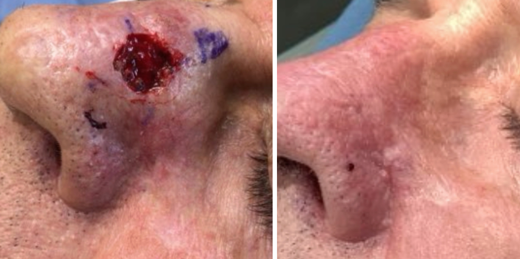
Defects of the nose can be difficult to repair because the nasal skin is relatively thick, has limited laxity, and the nose contains several distinct cosmetic subunits that can be distorted if tension is not carefully managed. Even small amounts of tissue movement can alter the natural contour of the nose, create asymmetry, or distort nearby structures such as the nasal tip, alar rim, or nostril. Careful reconstruction is therefore required to preserve both function and appearance.
This defect was repaired using an O-to-L advancement flap, a technique that recruits adjacent tissue while minimizing distortion of the surrounding nasal anatomy. This approach allows the wound to be closed with a good match in skin color and texture while maintaining the natural contours of the nose.

The area where the ear attaches to the side of the head can be particularly challenging to reconstruct because of the limited tissue laxity, the natural crease between the ear and scalp, and the need to preserve the ear’s position against the head. In this case, the defect was repaired with a rhombic transposition flap, which allowed adjacent skin to be recruited and rotated into the defect while distributing tension over a broader area. This technique helped preserve the natural contour where the ear meets the scalp, maintain normal ear positioning, and achieve an excellent cosmetic and functional outcome.
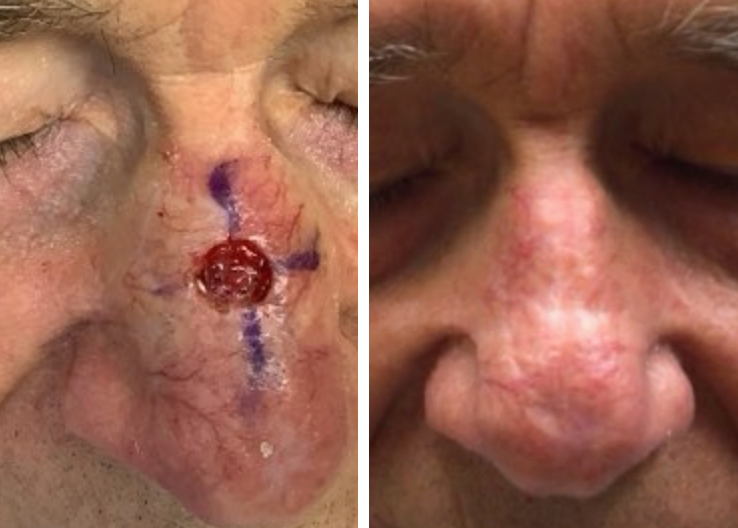
The nasal dorsum occupies a prominent position in the center of the face, making reconstruction particularly important from both a functional and cosmetic standpoint. Because the skin over the bridge of the nose is relatively inelastic and closely adherent to the underlying structures, surgical repairs must be carefully planned to avoid creating contour irregularities, visible asymmetry, or unnatural changes in the shape of the nose. Achieving a successful outcome requires a balance between complete cancer removal and preservation of the nose’s natural appearance.
Reconstruction was performed with a bilateral advancement flap. This technique utilizes adjacent skin from both sides of the defect, allowing the wound to be closed in a controlled and balanced fashion. By advancing tissue toward the center from opposite directions, tension is dispersed more evenly, and the normal contours of the nasal bridge are better maintained. The flap also provides an excellent match in skin thickness, color, and texture, helping the repair blend naturally with the surrounding tissue.
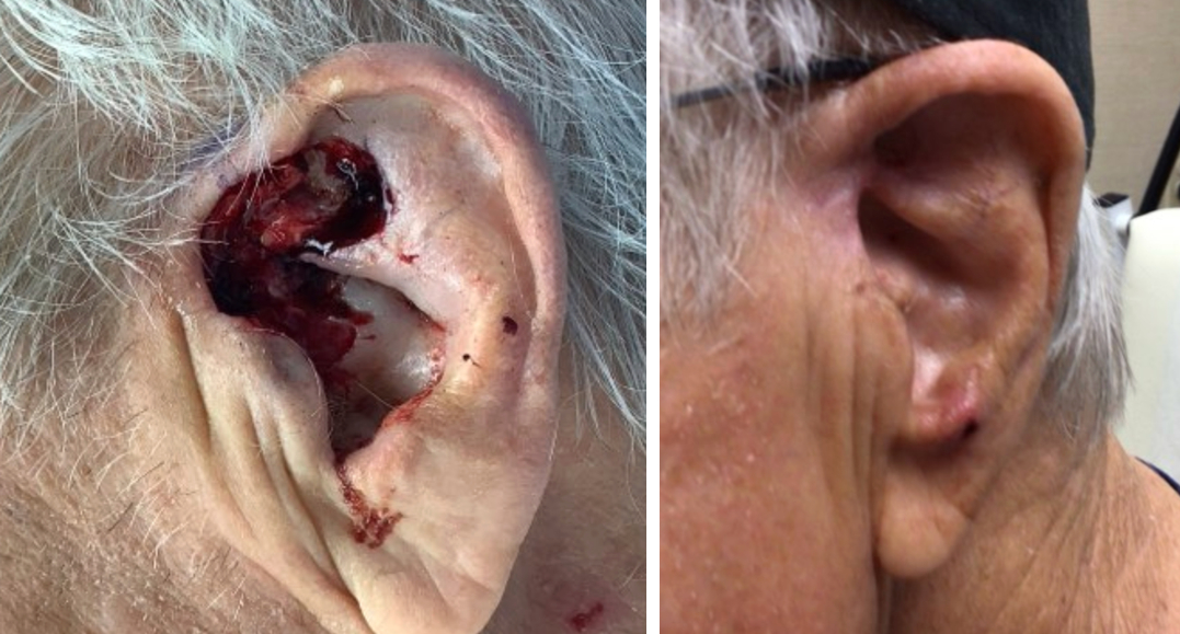
The ear contains complex contours that contribute to its natural appearance and function. Reconstruction of defects in this area can be particularly challenging because the skin is thin, closely associated with the underlying cartilage, and there is limited adjacent tissue available for closure. In this case, a very large infiltrative basal cell carcinoma was removed. Due to the extensive subclinical spread of the tumor, multiple stages of Mohs micrographic surgery were required to achieve complete cancer clearance, resulting in a substantial surgical defect.
Given the size of the defect, reconstruction was performed using a full-thickness skin graft harvested from the skin near the clavicle. This technique allows healthy skin to be transferred to the defect while preserving the natural contours of the ear. Skin from the clavicular region provides a suitable donor source with favorable color and texture characteristics. The graft was carefully secured into the surgical site to promote optimal healing, preserve the ear’s architecture, and achieve the best possible functional and cosmetic outcome.